When can I start exercising a dog after a muscle strain?
15 Sep, 2020
When our dogs injure a muscle, however slight, we typically give them a couple of days rest and then allow them to return to normal exercise. Is this approach sufficient to restore the dog’s muscle strength or are we risking re-injury?
What is muscle injury?
Muscle injury involves tearing of muscle fibres and disruption of vascular and connective tissue support. The most severe muscle injury will involve a complete rupture of muscle fibres across the muscle belly. In severe cases, the dog will show very obvious signs of injury including an inability to weight bear on a limb, loss of function, and signs of pain such as extreme sensitivity to touch or vocalising.
At the other end of the scale, less severe injuries may only affect only a few muscle fibres. With minor muscle injuries, we may not see any outward sign of the injury. The dog may initially limb for a few steps and then show no other signs of an injury.
What are the common causes of muscle injuries in dogs?
Muscle injuries can be caused by a range of incidents and actions including:
- Laceration or puncture where the skin is cut or torn and the underlying muscle is damaged.
- Contusion such as when the dog contacts an object or the ground, as in the case of a slip or car accident.
- Forceful muscle contraction like when the dog props, turns quickly or performs a sudden action.
What are the stages of muscle healing in dogs?
There are several phases of healing that occur, regardless of the cause or severity of the injury.
Inflammation phase
The inflammation phase is sometimes referred to as the “acute stage”. The inflammation phase occurs in the first 72 hours after injury. This phase is characterised by the following:
- Accumulation of fibrin (blood clotting agent) at the site of the injury which forms a hematoma (bruising, blood clotting).
- Inflammatory immune response with production of neutrophil and macrophage (M1) to remove necrotic debris at the site of the muscle fibre rupture.
- Restoration of vascular supply to the injured muscle fibres with capillary regrowth.
- Accumulation of substances such as fibronectin. Fibronectin binds extracellular components of the muscle including collagen and fibrin. The fibrin – fibronectin network forms the scaffold upon which platelets and fibroblasts adhere. Platelets and fibroblasts are the cells that hold tissues together and produce an extracellular matrix upon which the muscle fibres can regenerate during the repair phase.
Repair phase
The repair phase starts from three (3) days to up to six (6) weeks after injury. During this phase, there is competition for regenerating functional muscle fibres and producing fibrous scar tissue. In the repair phase, the extracellular matrix comprised of fibroblasts including collagen is produced. This extracellular matrix provides a structure upon which muscle fibres (created by myoblasts) regenerate at the injury site.
When there is inadequate:
- source of myoblasts (substrate for production of muscle fibres),
- vascularisation (blood flow),
- innervation
- or excessive stress across injury site that creates a gap
then excessive collagen is deposited and fibroblastic activity occurs. This leads to excess fibrous scar tissue forming at the injury site. This fibrous tissue forms a barrier to regenerating muscle fibres crossing the injury site and prevents the muscle from returning to its pre-injury tensile strength. In this state, the risk of re-injury is high.
Remodelling phase
The remodelling phase occurs 6 – 12 weeks after the injury. During this phase, normal tissue regeneration required for muscle function occurs. The ratio of functional muscle fibres and scar tissue depends on the size of the gap as well as the level of mobilisation and stress at the injury site.
How to restore muscle function and strength?
The type of healing (excessive scar tissue vs regenerated muscle fibres) can be influenced by applying appropriate stress and motion at the injury site during the appropriate healing phase. An animal study concluded that introducing controlled mobilisation after 3 – 5 days of immobilisation (directly following the muscle injury) had the following positive effects:
- accelerates the generation of muscle fibres,
- reduces connective scar tissue
- accelerates capillary growth
- increases tensile properties of the muscle
In a study of rats, the researchers induced a partial muscle tear to the gastrocnemius muscle to test the effect of rest and controlled mobilisation on restoring muscle strength. The study involved 132 rats that were split into the following four groups:
- No treatment group – After the injury, these rats were returned to their cages and allowed to move freely.
- Immobilisation group – After the injury, this group had a plaster applied to the injured leg so it was completely immobilised for 21 days.
- Immobilised for 2 days group – After the injury, this group had a plaster applied for two days. After this time, the rats were exposed to controlled exercise on a treadmill. Initially, the treadmill sessions were 20 minutes twice a day. Over the course of the study, the exercise sessions were gradually increased to a once daily 60 minute session.
- Immobilised for 5 days group – This group were treated the same way as the above the group, except the injured leg was immobilised for five (5) days.
At day 2 post injury, while still in the inflammation phase of healing, hematoma and disrupted muscle fibres were observed in the not treatment and immobilised group. In these groups, inflammatory cells were present around the damaged fibres, this was particularly the case in the no treatment group.
In the no treatment group, fibronectin was present along strands of fibrin indicating the formation of a matrix onto which fibroblasts can anchor. In the immobilised group, fibronectin was present throughout the injury site indicating that a scaffold for muscle fibres had not yet started to form. Early in the inflammation phase, however, the intensity of fibronectin in the not treatment group began to decrease. This finding indicates that the resumption of normal activity acted as a pump to remove the fluid containing fibronectin from the injury site. This conclusion may also explain the presence of high levels of fibronectin in the immobilised group for several days post injury.
During the repair phase, differences in the recovery protocols were becoming obvious. In the immobilised group by day 5 post surgery, the hematoma had almost resolved itself. This was also the case by day 7 in the group that was immobilised for 5 days. This finding indicates that immobilisation post injury is effective in reducing swelling around a muscle injury.
Later in the repair phase, by day 21 there was evidence in all groups of muscle fibres regenerating in the scar tissue. In the group that was immobilised for 5 days before commencing controlled exercise, the orientation of the regenerated muscle fibres was less interlaced than the other groups and more parallel. Parallel muscle fibres have greater tensile strength as they contract along the same axis.
By day 56, in the remodelling phase, there was evidence of fibrous scar tissue in all groups. The most significant amount of scar tissue was observed in the immobilised for 2 days group. In this group and the immobilised group, the regenerated muscle fibres were smaller compared with muscle fibres in the uninjured area. These findings indicate that returning to activity too soon, risks re-rupturing injured muscle fibres resulting in a larger injury area. It also suggests that long term immobilisation and returning to activity too soon, impacts the regeneration of muscle fibres.
At day 56, the regenerated muscle fibres in the immobilised for 5 days group were largely parallel indicating a restoration of muscle function and strength.
What are the effects of physical activity on muscle healing?
In the group that was immobilised for 5 days before starting a controlled exercise programme, there was an accelerated appearance of Type I collagen. Type I collagen appears after Type III collagen and it forms thicker, stronger fibres than Type III. This finding indicates that immobilisation may allow the muscle to gain sufficient strength to reduce the risk of re-rupture. By day 7, after two days of controlled exercise, the subjects in this group showed no signs of re-rupture.
In the group that was immobilised for two days before resuming controlled exercise, a fibrin clot containing fibronectin was detected at day 7, after 5 days of controlled exercise. This finding indicates that the muscle was not strong enough for the stresses of exercise due to a lack of connective tissue. The result was the formation of a larger area of granulation tissue and the ongoing presence of a larger fibrin-fibronectin clot to trap fibroblasts into the area of damage.
This study showed that different periods of immobilisation affected the remodelling of injured muscle and the resorption of connective tissue. In the immobilised for 5 days group, the connective tissue was resorbed by the 8 week follow up point. While in the immobilised for 2 days group, a fatty necrosis (dying cells) at the centre of the injury site was still present three (3) weeks post injury. At 8 weeks after the injury, the deposition of connective tissue was still present in this group, as a result of re-rupturing the injury site. Re-injuring the pre-existing muscle lead to additional extracellular matrix being deposited within the pre-existing, mature connective tissue. Poor resorption of the connective tissue prevents the regenerated muscle fibres full penetrating the connective tissue which affects muscle strength.
Takeaway messages for dog owners
This study provides the following insights for dog owners rehabilitating their dog after a mild to moderate muscle injury, that does not involve breaking the skin.
- Immobilisation for 5 days post injury – For dogs, immobilisation may involve bracing the affected limb to prevent movement or confinement to their crate for five days after an injury.
- Return to controlled exercise – In this study, exercise was slowly re-introduced. On the first day, the rats performed two (2) 20 minutes sessions on the treadmill. It should be noted that these animals were conditioned to treadmill running before their injury. For dogs, controlled return to exercise should consider the following:
- Performing exercise to which the dog was conditioned prior to the injury e.g. lead walking, swimming, underwater treadmill etc
- Several shorter sessions is more beneficial than one long session.
- Ensure the only exercise the dog undertakes is the controlled, supervised sessions. Between sessions, the dog should be crated or their movement restricted.
- Increase the duration and intensity of exercise gradually, over the course of several weeks.
Full Stride provides remedial canine massage treatments to keep dogs active and moving without pain. Treatments are offered at my home based clinic on Brisbane’s northside. Home visits are also available within a service area.
Until next time, enjoy your dogs.
Sources:
Lehto, M., Duance, V. C., & Restall, D. (1985). Collagen and fibronectin in a healing skeletal muscle injury. An immunohistological study of the effects of physical activity on the repair of injured gastrocnemius muscle in the rat. Bone & Joint Journal, 67(5), 820-828.
Waters-Banker, Christine (2013) “Immunomodulatory effects of massage in skeletal muscle”, Theses and Dissertations – Rehabilitation Sciences, Paper 18
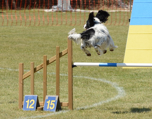
Image by SnottyBoggins from Pixabay